DESCRIPTION
mCherry is the second generation monomeric red fluorescent protein that was derived from proteins originally isolated from Cnidarians (jelly fish, sea anemones and corals), such as GFP or DsRed. mCherry is widely used as a fluorescent tracer in transfection and transgenic experiments because of its better performance in brightness, photostability and monomeric structure. Since mCherry requires no additional substrates or cofactors, mCherry can be easily detected under a fluorescence microscope.
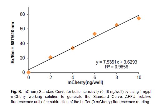
However, most imaging studies of mCherry are only qualitative. BioVision’s mCherry Quantification Kit provides an easy 96 micro-plate assay to analyze mCherry expression level in cells or tissues quantitatively. Cells or tissues can be homogenized directly in the mCherry Assay Buffer. The amount of mCherry is determined by comparing its fluorescence (Ex/Em= 587/610 nm) with a mCherry standard that is provided in this kit. Each kit provides sufficient reagents to perform up to 100 assays.
mCherry Quantification Kit
IHC using an anti-mCherry antibody (fluorescent and stained image). Intestinal tissue from a transgenic mouse expressing mCherry in all tissues. (A) Fluorescent image detecting mCherry expression and (B) HRP-stained image. Following isolation and fixation in 4% paraformaldehyde, mCherry Monoclonal Antibody (16D7) (Cat. No. M11217) was used at a 1:15,000 dilution. The ImmPRESS™ Anti-Rat Ig (peroxidase) Polymer Detection Kit (Vector Laboratories) was used according to manufacturer’s instructions; sections were incubated in peroxidase substrate solution until the desired stain intensity developed.
Flow cytometry analysis of cells expressing mCherry. U20S cells expressing mCherry were analyzed using 405 nm excitation and 450/40 nm band pass emission on an Attune Acoustic Focusing Cytometer. The histogram shows cells stained with mCherry rat monoclonal antibody conjugated with Pacific Blue (black line) and unstained cells (gray line).
| Cat # +Size | K971-100 |
|---|---|
| Size | 100 assays |
| Detection Method | Fluorescence (Ex/Em 587/610 nm) |
| Species Reactivity | Mammalian |
| Applications | It detects a wide range of mCherry (0.01-1 ng/µl) |
| Features & Benefits | • Simple procedure; takes ~ 20 minutes • Fast and convenient |
| Kit Components | • mCherry Assay Buffer • mCherry Standard |
| Storage Conditions | -20°C |
| Shipping Conditions | Gel Pack |
| USAGE | For Research Use Only! Not For Use in Humans. |